
Careers and study
2025-2026 Hector Maclean Scholarship Projects
Explore research projects especially tailored for the Hector Maclean Summer Scholarships and find out how to apply.
The Hector Maclean Scholarship funds summer research studentships within the Department of Ophthalmology, based at the Centre for Eye Research Australia (CERA) and The Royal Victorian Eye and Ear Hospital.
Eligibility
Students from The University of Melbourne science, biomedicine, medicine or other relevant undergraduate and postgraduate coursework degrees are eligible to apply. Please note students enrolled at other universities cannot be considered. This program is not for credit and is not examined. Selection for the Studentships is based on academic merit.
Duration
4-8 weeks, full-time or part-time, dependent on the supervisor’s approval. Positions can run across Block 1 (3 November to 19 December 2025) or Block 2 (12 January to 27 February 2026), with exact dates to be negotiated between the student and supervisor.
Scholarship value
$200 per week, pro-rata, tax-free, up to a maximum of $1600.
Explore our projects below and apply
Learn more about the four projects tailored for students applying for Hector Maclean Summer Scholarships and find out how to apply.
If you have any questions about our research projects or the application process, please contact: education@cera.org.au
Please note: Applications close 5 pm Sunday 5 October 2025.
Reducing blindness from Fuchs’ endothelial corneal dystrophy (FECD)
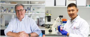
Supervisors: Professor Mark Daniell and Dr Gink Yang
Research Group: Corneal research
Timeline: Flexible (to be negotiated)
Selection criteria: Biotechnology or Biomedical science students who are interested in drug development and validation. The successful applicant will possess laboratory experience in molecular biology and tissue culture and be able to work independently. Minimal WAM = 75.
About this project:
Fuchs’ Endothelial Corneal Dystrophy (FECD), a blinding disease remains the most common indication for corneal transplantation in high-income nations.
To develop less invasive therapies and reduce the need for surgery for early symptomatic patients, Next Generation Sequencing including DNA and RNA based approaches has been used to reveal a range of therapeutic targets.
Our latest snRNA-sequencing study investigated the spatial expression of FECD-related genes at a single cell level using FECD patient endothelium and healthy human cornea as control. At the ligand level, high levels of FGF7, FGF9 and FGF10 expression was found in the corneal endothelium when compared to other corneal layers and ocular tissues in this study. Specifically, FGF7 and its receptors FGFR1 & FGFR2 are highly expressed in FECD endothelium compared to healthy controls.
The current project aims to investigate if the FGF7 expression in a protein form is consistent with our preliminary scRNA-seq data on discarded FECD patient tissue following corneal transplantation surgery. Results from this study will be steppingstones for the investigation of FGF7 as a potential therapeutic target in treating FECD.
Questions?
If you have any questions about our research projects, please don’t hesitate to contact: education@cera.org.au
Low-vision functional analysis

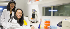
Supervisors: Dr Carla Abbott, Associate Professor Penelope Allen and Dr Myra McGuinness
Research Groups: Bionic Eye and Biostatistics research teams
Timeline: 4-8-week project to be conducted November-December 2025.
Selection criteria: No prior knowledge is required, but students with current skills in Heyex, Redcap and Stata will be at an advantage. Students must be prepared to travel to participants’ home locations for the functional assessments.
About this project:
Improving functional outcomes for people with low vision is important to their independence and quality of life. Our team works with participants in low-vision clinical trials to help improve functional outcomes.
The project involves two streams of work:
- Helping to collect and analyse data in the local environs of people with low vision to assess a novel tool we have created regarding how they use their different senses to approach tasks of daily living.
- Conducting image analysis in a subgroup of six participants who currently have a bionic eye implanted to measure the long-term tolerability of the bionic prosthesis by the eye. The retinal imaging data has already been collected, and the student will follow our established procedures to perform the analysis.
Students will learn skills in orientation and mobility assessments in home and local environments, image analysis skills in Heyex retinal imaging software, use a Redcap database to capture results and use Stata software to analyse the outcomes.
Questions?
If you have any questions about our research projects, please don’t hesitate to contact: education@cera.org.au
Scarring the vision: Unraveling fibrotic pathways after glaucoma filtration surgery
Supervisors: Dr Jennifer Fan Gaskin and Dr Elsa Chan
Research Group: Ocular fibrosis research
Timeline: Flexible (to be negotiated)
Selection criteria: No prior experience is required.
About this project:
Glaucoma filtration surgery is the gold standard glaucoma operation but also the last option for treating glaucoma when other therapies such as medications or lasers are not sufficient.
During the surgery, a new drainage pathway is created by which aqueous fluid inside the eye can escape, thereby lowering the intraocular pressure. Scarring occurs as a result of a natural wound healing response, however, uncontrolled scarring can block this new drain. Chronic use of glaucoma eyedrops, which is common amongst glaucoma patients, can also predispose to scarring following glaucoma surgery.
Minimisation of scarring is currently treated through the application of cytotoxic agents such as Mitomycin C (MMC) during the surgery, nevertheless scarring remains a problem in some patients. Moreover, MMC is an anti-cancer drug that can lead to severe complications and even blindness. Due to these considerable risks, it is not uncommon to only perform trabeculectomy when patients have started to lose vision.
There is an urgent need to improve the long-term efficacy and safety of glaucoma surgeries. Our unit explores the underlying fibrotic pathways with the aim of developing new strategies to improve current anti-scarring therapies.
Students would have the opportunity to learn and develop a number of techniques including cell culture and ELISA.
Questions?
If you have any questions about our research projects, please don’t hesitate to contact: education@cera.org.au
Quantifying interpericyte tunnelling nanotubes in the optic nerve

Supervisor: Dr Anna Wang
Research group: Visual Neurovascular Research
Timeline: 4-week project to be conducted November-December 2025.
Selection criteria: No prior experience is required.
About this project:
Glaucoma is the leading cause of irreversible blindness worldwide. It is characterised by death of retinal ganglion cells whose axons make up the optic nerve. Elevated intraocular pressure is a major risk factor, yet despite interventions to lower it, approximately 25–45 per cent of glaucoma cases continue to progress. The mechanisms underlying RGC death in glaucoma remain incompletely understood.
The vascular theory of glaucoma proposes that inadequate blood supply contributes to retinal ganglion cell axon loss. Neurons, especially retinal ganglion cells, have a high metabolic rate and must communicate with blood vessels to secure adequate oxygen and nutrients. Disruptions to the blood supply can, therefore, lead to neuronal dysfunction. Although the vascular supply of the optic nerve is critical for axonal health, it remains poorly defined.
Recently, our lab has identified pericytes as important mediators of precise co-ordination of blood flow. These cells wrap around blood vessels, and some are connected by fine processes called interpericyte tunnelling nanotubes (IPTNTs). Importantly, IPTNTs co-ordinate the dilation and constriction of distinct capillaries in response to retinal neuronal activity.
This project aims to examine IPTNTs in the optic nerve and whether they are damaged in the optic nerve of a mouse glaucoma model. Students will learn how to analyse confocal images, perform immunohistochemistry, and mount tissue samples for imaging.
Questions?
If you have any questions about our research projects, please don’t hesitate to contact: education@cera.org.au