2026 Gerard Crock Lecture: Professor Pete Williams
Video
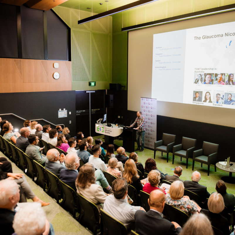
2026 Gerard Crock Lecture: Professor Pete Williams
Watch Professor Pete Williams present the 17th Gerard Crock Lecture: “From bench-to-bedside (and back again): Protecting and repairing the retina and optic nerve in glaucoma.”
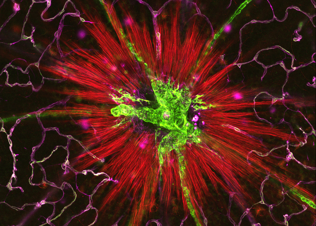
Glaucoma is a progressive neurodegenerative disease that leads to the loss of retinal ganglion cells in the eye and irreversible vision impairment.
For the 17th Annual Gerard Crock Lecture, Professor Pete Williams presents new research targeting NAD metabolism for neuroprotection. NAD is a coenzyme found in all living cells, which is essential for energy production.
Pete’s research highlights how restoring energy levels in the eye can protect retinal cells and preserve vision. He discusses how this work, complemented by ongoing clinical trials and drug development, aims to enhance the resilience of neurons and support new treatments that go beyond just lowering pressure in the eye.
Pete Williams joined CERA in January 2026 as a Research Lead, establishing the new Neuroprotection and Repair Unit. He comes to CERA from the Karolinska Institutet, Sweden, where he was the Ulla and Ingemar Dahlberg Professor in Vision Science.
His new research group at CERA explores how bioenergetic insufficiency drives neurodegeneration in conditions such as glaucoma and is developing new treatments based on these findings.
He is a renowned neuroscientist with research spanning from the laboratory through to clinical trials, and was the first to show the neuroprotective potential of nicotinamide in glaucoma.
This has now led to multiple clinical trials across the globe to validate nicotinamide as a potential new treatment for glaucoma. The Gerard Crock Lecture series honours the memory of renowned ophthalmologist, the late Professor Gerard Crock, inaugural Ringland Anderson Professor of Ophthalmology at the University of Melbourne.